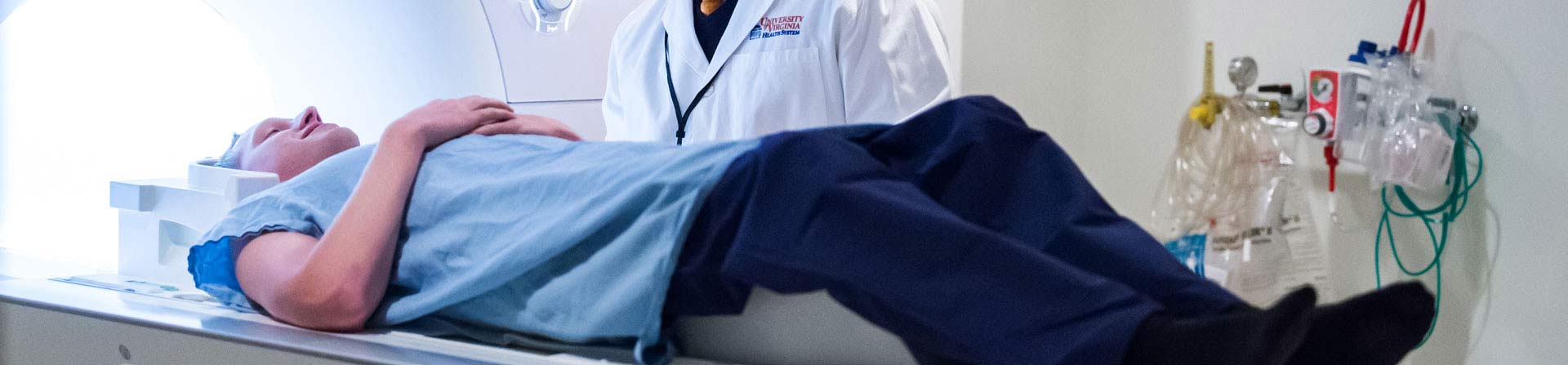
Neuroradiologists play a key role in diagnosing and treating neurological conditions. They use high-tech imaging that focuses on the brain, spine, head and neck.
At UVA, our teams of highly trained specialists work together, aiming for the best outcomes possible.
Our diagnostic neuroradiologists and interventional neuroradiologists conduct research that keeps them at the forefront of the field. We're constantly improving our processes and enhancing the precision of our tools.
The Crucial Role of Imaging
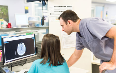
Diagnostic neuroradiologists examine brain scans to detect and diagnose conditions such as Parkinson's disease, tumors, aneurysms and vascular malformations.
But our work doesn't just find serious disease or problems in the brain. We provide detailed images to help your neurosurgeon understand treatment options. While often behind the scenes, neuroradiologists play an essential part in neurosurgery and neurology procedures.Guided by accurate, detailed scans, your healthcare team can plan a successful treatment.
Imaging Tests We Use
- Angiography/Neuroangiography – This test is used to find and map blood vessels in the brain. After injection of a dye into a deep artery, X-rays follow its flow throughout the blood vessels of the brain. Angiography helps to make a diagnosis of stroke, aneurysm, arteriovenous malformation (AVM), tumor, clotting, and arterial stenosis.
- Biplane angiography – This test creates 3-D brain images. It is used in patients who have aneurysms, arteriovenous malformations (AVMs), and other conditions. We use it to find the exact location of an issue in your brain before surgery.
- Computed tomography (CT) – Uses an advanced X-ray machine and a computer to create a detailed picture of the body’s tissues and structures. It can help us determine the location and cause of a problem. It can also help detect swelling and bleeding.
- Electroencephalogram (EEG) – Measures and records the electrical activity of your brain using sensors (electrodes) attached to your head.
- Electromyography (EMG) – Measures how muscles and nerves respond to electrical activity. It helps diagnose muscle conditions that can cause muscle weakness, like muscular dystrophy and nerve disorders.
- Magnetic resonance imaging (MRI) – Uses a magnetic field to show healthy and diseased tissue. MRI is better than CT for areas located near bone. It also provides a 3-D image of your condition.
- Functional magnetic resonance imaging (fMRI) – Clinical fMRI brain-mapping technology can be used before and during surgery. It also helps us evaluate neurological issues.
Online Imaging Results
You can find, save, print, and share your radiology results and scans using the secure online platform, MyChart.
A New Kind of Surgery
If you're facing brain or spine surgery, you could benefit from an interventional neuroradiologist.
Interventional neuroradiologists perform minimally invasive procedures on the veins and arteries within the brain or spine. Sometimes called endovascular neurosurgeons, they provide precise, image-guided treatments that replace traditional surgery.
These kinds of intervention include:
- Endovascular embolization, which controls bleeding in the brain and prevents rupture
- Endovascular coiling, which uses a catheter
See maps, contact info for the Interventional Neuroradiology clinic.