Heart Imaging
Make an Appointment
For the Charlottesville area:
For Northern Virginia:
If you're concerned about a heart problem or vascular disease, finding the right diagnosis as early as possible gives you the best chance for restoring a healthy heart.
At UVA, we're using imaging technology to better diagnose and treat heart and vascular conditions. New imaging tools help your care team understand your condition and play a key role in deciding whether we do a procedure in the first place.
And, as a leading research institution, we're always working on moving safer, more effective heart imaging from the lab into the exam room.
Heart Imaging and Screening Options
We offer a full range of the most advanced imaging and screening tests — many of which use less invasive approaches and cause little or no pain. These include:
Ankle-Brachial Index
The ankle-brachial index is a non-invasive screening that measures blood pressure in a patient's arms and legs to diagnose peripheral arterial disease (PAD).
Cardiac Catheterization
Cardiac catheterization evaluates blood flow and pressure to the arteries that feed the heart (coronary arteries). Your doctor will insert a long, thin tube (catheter) into a blood vessel in your arm, upper thigh or neck and guide it to your heart to detect a variety of problems in the heart, including narrowing or blockages of arteries and poor heart valve function.
For more information, call our Cardiac Catheterization Lab at the Heart & Vascular Center.
Cardiac CT
Cardiac CT uses an X-ray to take a detailed picture of the hear to reveal potential problems such as coronary artery disease, aneurysms, embolisms and calcium buildup in the arteries.
Cardiac Monitoring
Cardiac monitoring helps doctors diagnose heart rhythm disorders and determine appropriate treatment options. The Holter monitor (worn for 24 hours) or the event monitor (worn for a month) may detect episodes of irregular heartbeat. Another monitoring device, the loop recorder, may be implanted and used to record your heart’s rhythm for up to three years.
Cardiac MRI
Cardiac MRI radio waves produce a computerized image of your heart and blood vessels. The non-invasive test helps diagnose and assess coronary artery disease, congenital heart disorders and determine the cause of heart failure, among other issues.
Echocardiogram
An echocardiogram uses ultrasound waves to take detailed pictures of your heart, which allows doctors to evaluate heart muscle function and structure. We offer transthoracic echocardiogram (TTE), stress echoes and transesophageal echocardiogram (TEE).
Learn more about echocardiography.
Genetic Testing
Most patients are diagnosed by ECG or EKG, but other tests like MRIs and Holter monitoring may also be used. Genetic testing can help determine which family members are at risk for conditions like hypertrophic cardiomyopathy. An early diagnosis can prevent serious complications like sudden cardiac arrest and heart failure.
Learn more about our genetics program.
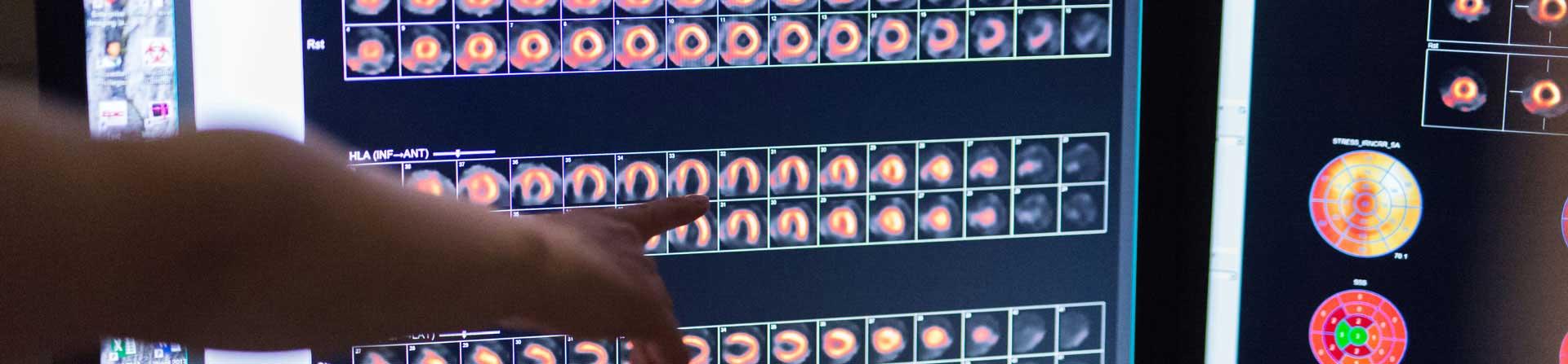
Nuclear Imaging
Nuclear imaging uses SPECT (single photon emission computerized tomography) containing very low levels of radioactivity to produce images that identify decreased areas of blood flow. Our imaging specialists also use cardiac PET, a newer nuclear technology that can precisely measure blood flow and identify disease in the small blood vessels supplying the heart.
Learn more about cardiac PET.
Vascular Ultrasound
Specially trained vascular technologists and sonographers diagnose carotid stenosis with a vascular ultrasound. Vascular stenosis is a risk factor for stroke. Vascular ultrasound is also used to screen for an aortic aneurysm.
Expertise in Imaging
Cardiologist Chad Hoyt, MD, discusses advanced imaging options at UVA.
My name is Chad Hoyt. I'm a clinical cardiologist here at UVA Health, and I'm also the Executive Director of Clinical Growth and Outreach. At UVA Health, we have an incredible imaging department, probably world-renowned, so we offer CTA of the coronary arteries. That's a CAT scan procedure. We offer MRI of the heart. We offer PET scanning of the heart and all of the different ultrasound imaging modalities.
The tremendous benefit to patients for advanced imaging is that we can detect disease at a much earlier stage and we can detect it noninvasively, so there's less risk to the patient. The newer imaging procedures have less radiation than some of the older machines. Some have asked, why do we call it advanced imaging? I would say that it's primarily because these machines are very, very technologically sophisticated, they're expensive, and so it involves a lot of specialized physician expertise, fellowship expertise in order to read them, so it's not a run-of-the-mill EKG or chest x-ray. They take time to do and they're at a significant expense, but they give us amazing information and detail.
Patients often ask if they can refer themselves for an advanced imaging test, and the answer to that is that you really need a referral from a physician because some of the imaging procedures involve some radiation exposure and you need to be informed and we need to make sure that you have a good indication for the test.
I would really encourage patients to come to UVA Health for heart imaging because we have a world-renowned faculty, we have an incredible imaging research department, we have great imaging technology and equipment, and I think it's the best in Virginia, some of the best in the East Coast.
UVA Health Doctors: Leaders in Heart Imaging
It’s not enough to have the right test. You need the right people to interpret the results.
Our doctors are experts in their imaging fields. Many serve in leadership roles in national imaging societies. This means UVA doctors aren't just following national guidelines and best practices. In many cases, they're the ones who are trusted to write them.
Our doctors also know the importance of matching the right test to the right patient, and will only ask for tests they believe are absolutely necessary.