Imaging & Radiology

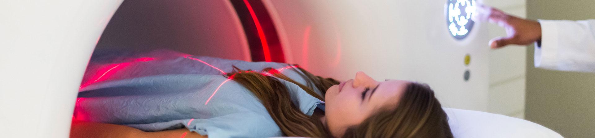
patient getting scanned

Find high-quality scans from caring professionals. We offer personalized, subspecialty imaging services, from routine X-rays to highly advanced radiology treatments.

Imaging at UVA Health: The Advantages

The expertise of both our board-certified radiologists and experienced technologists runs the gamut of imaging care. Along with consulting with referring doctors, we continue to push the boundaries of research. We're always finding new ways to use imaging to advance the possibilities of patient care.

At UVA Health, you'll benefit from:

  • Knowledgeable staff with extensive training and experience
  • Images of the highest quality diagnostic
  • Expert radiologists who produce more accurate reports and have higher detection rates
  • State-of-the-art equipment, including one of the industry’s most comfortable and accurate MRI machines
  • Minimally invasive treatments that can replace traditional surgery

Preparing for Your Scan

General guidelines for your appointment:

  • All exams require a doctor's order.
  • We don't schedule X-rays, but offer them on a walk-in basis only.
  • Leave jewelry and other valuables at home.
  • Bring a driver's license and insurance card (if you have one).
  • Be sure to arrive at least 15 minutes early.

Get a Scan Near You

You can make an appointment for a mammogram or other routine screening at a number of convenient locations.

Services We Offer

See your scans

See Your Scans

After your scan, you can see the results in MyChart. This secure, free portal lets you save, print, and take your scans anywhere.

Radiology & Imaging Trials
Research study for women scheduled for screening mammogram

The department of Radiology, Breast Imaging division seeks women ages 45-74 that are due for their annual screening mammogram for a research study. The purpose of the study is to help us develop strategies that tailor breast cancer screening to the risks of individual women You may be eligible for this study if you have no symptoms of breast disease, no history of breast cancer and currently do not have breast implants or other breast enhancements If you decide to participate, you will be involved in this research study for up to 8 years starting with your first mammogram. You will be randomized (assigned by a computer) to get either 3D or 2D mammograms for the first 5 years of the study. We will follow your breast cancer status for up to 8 years in total, including the screening years, by reviewing your medical records and, if needed, contacting you by phone Participant’s insurance company will be billed for mammogram
Research Study for Active-Duty Service Members and Veterans with a History of Blast Exposure

The Department of Radiology and Medical Imaging seeks active-duty service members and veterans aged 18-65 with a history of blast exposure for a research study. The purpose of the study is to better understand brain injury in people who have been exposed to blast events. You may be eligible for this study if: you are an active-duty service member or veteran, have a history of blast exposure and are willing to travel to Charlottesville, VA. Study involves completing assessments and questionnaires, an MRI scan, a PET-CT scan where you will be given a radiotracer and providing a blood sample. Study procedures will be completed in a one-day visit lasting approximately 6-8 hours. Study-related procedures provided at no cost.

Breast cancer screening study: Contrast Mammography Imaging Screening Trial (CMIST) Research Study for women with dense breast tissue stated on a current mammogram report and are scheduled or due for an annual screening mammogram.

The Contrast Mammography Imaging Screening Trial, or CMIST is comparing two methods of mammography to help women and their doctors decide the best ways to screen for breast cancer in the future. The CMIST research study will follow women already planned to get three-dimensional (3D) tomosynthesis screening mammography routinely for breast cancer. The study compares two different types of mammograms used to screen for breast cancer in women who have breast tissue that appears dense on the mammogram. The two types of mammography methods being compared in CMIST are the three-dimensional (3D) tomosynthesis mammography that you are scheduled for and a contrast-enhanced spectral mammography. Our mammography clinic can provide both screening methods. Researchers want to find out which of the two approaches to breast cancer screening is better in the early detection of breast cancers for women with dense breast tissue. The department of Radiology, Breast Imaging division seeks women, ages 45-74 currently due for an annual screening mammogram for a research study. The purpose of the CMIST study is to compare two methods of mammography to help women and their doctors decide the best ways to screen for breast cancer in the future. You may be eligible for this study if: you have dense breast tissue as stated on previous mammogram report, are 45-74 years of age and have no history of breast cancer or breast implants. If you decide to take part in this study, you will be screened for breast cancer with both contrast-enhanced spectral mammography and digital breast tomosynthesis mammography when you enter the study. We will repeat those same tests in one year. Each visit could last up to 3 hours. Study-related contrast-enhanced spectral mammography provided at no cost. Participant’s insurance company will be billed for annual screening digital breast tomosynthesis (3D) mammography.

Research Study for Adults who are Healthy and/or E-cigarette user are invited to a Xenon MRI Imaging and Cardiopulmonary Stress Test

The UVa Health System, Department of Pulmonology, seeks healthy adult who are 18 to 35-years old that are e-cigarette users and non-users for a research study. The purpose of this study is to determine the changes to the lungs in people who uses e-cigarettes and whether a hyperpolarized gas called xenon-129 during MRI (Xe-MRI), can detect changes in lung function. The study will involve one study visit to determine your eligibility. Two study visits for an MRI with inhaled xenon gas and two visits for a cardiopulmonary stress test (CPET) over a one-year period. On the day of the Xe-MRI, computed tomography scan (CT) an electrocardiogram (EKG), PFT or breathing test, and a physical exam (including vital signs and listening to your heart and lungs) will be performed. Separate visits are required for a cardiopulmonary stress test. These tests let us know more about your lung function. If you qualify for the study, the study team will also obtain your blood and urine samples for biomarker studies, allowing the team to see what kinds of proteins are being produced by your body. Study can be completed in five visits that last about 3-4 hours. Study-related Xe-MRI, CT, CPET, EKG, PFT, blood and urine collection provided at no cost.

View All Clinical Trials
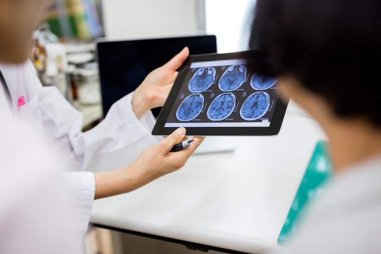
A doctor holds up a tablet with brain imaging visible

Work With Us

Find out more about available job openings on our imaging teams.